
Why does high myopia put you at risk of retinal detachment?
Written by Eagle Eye Centre
Published on April 7, 2026
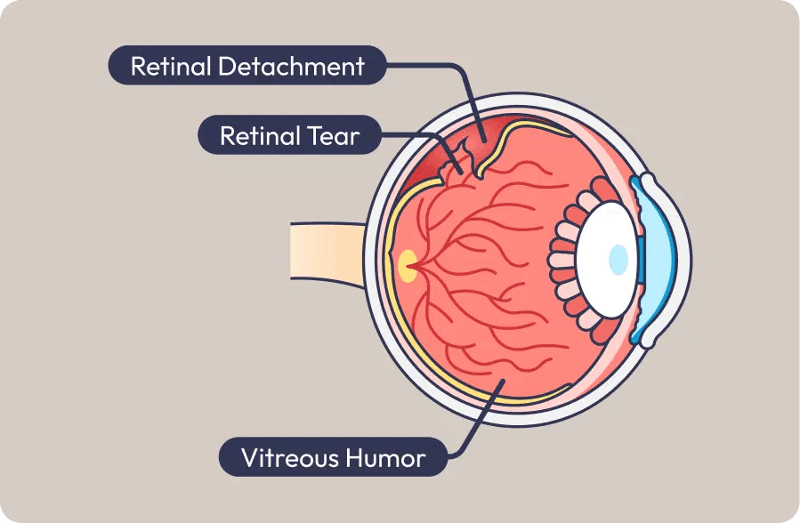
High myopia can increase the risk of retinal detachment, a sight-threatening eye condition that may require urgent treatment. In Singapore, where myopia is common, recognising the warning signs early is important to help protect vision.
This sudden and sight-threatening condition recently made headlines when Mediacorp actor Elvin Ng shared his emergency medical experience. On February 23, he noticed a dark, stationary shadow in the lower part of his right eye.
“When I moved my eye around, the shadow didn’t move. By the evening, the shadow had covered nearly half my eye and was near the centre.”
Suspecting it was related to his high myopia, he sought immediate medical attention and was diagnosed with retinal detachment, requiring emergency surgery on the same day.
Could this happen to you? Here is what to know about retinal detachment, why high myopia increases the risk, and the warning signs that should never be ignored.
What is the retina and what does it do?
The retina is a thin, light-sensitive layer of tissue lining the back of the eye. It plays a vital role in vision by receiving incoming light and converting it into electrical signals, which are then sent through the optic nerve to the brain. This is how you are able to see images clearly.
At the centre of the retina is the macula, which is responsible for sharp central vision and colour vision. The peripheral retina supports side vision and night vision. Because the retina is essential for clear sight, problems affecting the retina, including retinal tears and retinal detachment, can seriously threaten vision and require prompt medical attention.
What is retinal detachment?
The human eye is filled with a clear, jelly-like substance called the vitreous gel. As we age, this gel naturally liquefies, shrinks, and separates from the retina. However, if the gel pulls too hard, it can cause the retina to tear and eventually detach.
Why does high myopia increase the risk of retinal detachment?
High myopia increases the risk of retinal detachment because the eyeball is usually longer than normal. This extra elongation stretches the retina, making it thinner, more fragile, and more prone to developing retinal tears.
In adults, retinal detachment often begins with a retinal tear. When the retina is already weakened by high myopia, changes in the vitreous gel, minor eye trauma, or traction on the retina may increase the risk of a tear forming. Once a retinal tear develops, fluid can pass underneath the retina and cause it to detach.
This is why people with high myopia should take sudden eye symptoms seriously. If retinal detachment progresses and affects the macula, the part of the retina responsible for sharp central vision, urgent treatment or emergency surgery may be needed to help preserve sight.

Due to the high prevalence of myopia in Singapore, retinal tears and detachments are notably more common here compared to Caucasian populations. The incidence rate is between 10 to 20 cases per 100,000 people, and it remains a leading cause of sudden, permanent vision loss if not treated promptly.
Reference: HMI Medical News & Resources
Three retinal detachment warning signs
Recognising the early symptoms of a retinal tear is important. If a retinal tear is detected early during an eye examination, prompt laser treatment may help prevent it from progressing to retinal detachment. Seek immediate medical attention if you notice any of the following retinal detachment warning signs:
- Sudden floaters
Floaters may appear as a cloud of dust, cobwebs, specks, or moving shadows in your vision. When a retinal tear occurs, tiny blood vessels in the retina may bleed, releasing blood cells and pigment into the vitreous cavity. This can cause a sudden increase in floaters. - Flashes of light
Flashes of light may look like brief lightning streaks or flickers in your peripheral vision. These flashes can happen when the vitreous gel pulls on the retina and may be an early sign of a retinal tear or retinal detachment. - Sudden blurred or distorted vision
A dark shadow, grey curtain, or blocked area in your field of vision may suggest that the retina has started to detach. If retinal detachment spreads to the macula, it can cause severe blurring and loss of central vision.
Types of retinal detachment treatments
Once retinal detachment occurs, surgery is usually needed to reattach the retina, relieve traction on the retina, and help preserve vision. Two common retinal detachment treatments are vitrectomy and scleral buckle surgery.
- Vitrectomy
Vitrectomy is a retinal detachment surgery in which the surgeon removes the vitreous gel that is pulling on the retina. Scar tissue may also be removed, and the retina is gently repositioned. Retinal tears are then treated, and a gas bubble or silicone oil may be used to help keep the retina in place while it heals. - Scleral buckle
Scleral buckle surgery involves placing a flexible silicone band or sponge around the outside of the eye, known as the sclera. This gently pushes the wall of the eye inward, helping the detached retina move back into position and reducing the pulling force of the vitreous gel.
Treatment depends on whether there is a retinal tear (break) or a full retinal detachment. Our eye specialist will advise on the most appropriate treatment based on your condition.
Expert Q&A: Insights from EEC’s Senior Consultant Ophthalmologists
When it comes to retinal detachment, time is vision. Surgery must be performed urgently before the detachment reaches the macula (the central part of the retina responsible for sharp, detailed vision).
To understand how the surgery works and what to expect during recovery, our panel of Senior Consultant Ophthalmologists at Eagle Eye Centre: Dr. Stephen Teoh, Dr. Jacob Cheng, and Dr. Paul Zhao break down some of the most frequently asked questions
Q: How is retinal surgery performed? Is it done under local or general anaesthesia?
A: For straightforward cases requiring only a vitrectomy, our surgeons typically perform the procedure under local anaesthesia with sedation. However, for younger patients who may become restless under sedation and have a low risk for general anaesthesia (GA), we often prefer GA. If a scleral buckle (an external silicone band placed around the eye) is required, or if the surgery is expected to take longer, the procedure will also be performed under general anaesthesia.
Q: What is the purpose of injecting a gas bubble into the eye? How does it help reattach the retina?
A: A vitrectomy is the standard treatment for retinal detachment. At the end of the surgery, after the vitreous gel has been removed from the eye cavity, a gas bubble is injected. This bubble acts like an internal bandage—it floats upward and presses the detached retina flat against the wall of the eye, preventing fluid from entering through the retinal tear. Laser therapy is then applied around the edges of the tear to seal and reinforce it, allowing the retina to heal. The gas bubble is not permanent; the eye will naturally absorb it within 2 to 8 weeks, depending on the type of gas used.
Q: Why must patients maintain a face-down posture during their recovery? A: Patients with a gas bubble in their eye must maintain a strict face-down posture. Because gas naturally floats upwards, facing down ensures that the bubble accurately presses against the retina at the back of the eye. This posture also prevents the gas bubble from touching the lens at the front of the eye, which could otherwise cause the lens to become cloudy (leading to the formation of a cataract).
Q: Can retinal detachment happen again?
A: Yes, it can recur. This is typically due to the formation of new tears on the retina, or because scar tissue has developed and is pulling on the retina. This scarring condition is more commonly seen in younger patients.
Q: Is it safe to use ChatGPT or the internet to diagnose eye symptoms?
A: We are seeing an increasing number of patients using ChatGPT to self-diagnose symptoms like floaters. If this encourages patients to seek medical attention earlier and receive timely treatment, it is certainly beneficial. However, patients must never fully rely on all the information they get online or from artificial intelligence. Anyone experiencing visual symptoms must consult an eye specialist and undergo a comprehensive, dilated retinal examination. Only an accurate clinical diagnosis can ensure the proper management of your eye health.